
375A Concord Road, Concord West NSW 2138


PET Theranostics was referred a feline patient with a history of left-sided nasal adenocarcinoma that had been diagnosed approximately 12 months earlier. Following treatment with stereotactic radiation therapy (SRT) and carboplatin chemotherapy, previous CT imaging demonstrated a substantial reduction in tumour volume. However, a small contrast-enhancing nodule in the right ventral nasal cavity raised concerns about possible residual or recurrent disease.
In addition to the patient’s cancer history, clinicians were investigating a chronic liver disorder of unknown origin. Multiple diagnostic procedures, including ultrasound examinations, CT imaging and liver biopsies, had failed to establish a definitive diagnosis. PET-CT imaging was recommended to evaluate for active cancer, assess for metastatic disease, and further investigate the liver abnormalities.
Why is PET-CT recommended?
While conventional imaging can reveal structural changes, it may not always distinguish between active cancer, scar tissue, treatment-related changes or inflammation. PET-CT combines anatomical and metabolic imaging, allowing veterinarians to determine whether abnormalities exhibit increased metabolic activity, a feature typically associated with malignancy.
For this patient, PET-CT offered the opportunity to assess treatment response following radiation and chemotherapy while also evaluating the entire body for evidence of metastatic spread or other clinically significant disease.
PET-CT provided valuable reassurance that the patient’s previous nasal cancer treatment remained effective while helping identify other areas requiring ongoing monitoring.
Imaging procedure
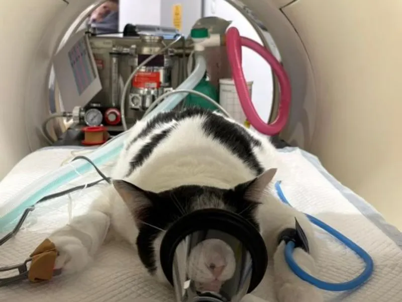
The patient underwent a whole-body PET-CT scan using F18-fluorodeoxyglucose (F18-FDG), a radiotracer that highlights areas of increased glucose metabolism. Imaging was performed approximately 60 minutes following intravenous administration of the tracer.
Physiological uptake was observed in expected tissues, including the brain, salivary glands, heart, abdominal organs and urinary system, confirming appropriate tracer distribution and scan quality.
Key findings
The most significant finding was the absence of evidence suggesting active recurrent nasal cancer. Although areas of turbinate destruction remained visible within the left nasal cavity, only minimal FDG uptake was identified, and no distinct mass effect was present. These findings were considered most consistent with post-radiation treatment changes rather than active malignancy.
Importantly, PET-CT also demonstrated no evidence of pulmonary metastasis. No pulmonary nodules were identified, and only mild enlargement of sternal lymph nodes with minimal metabolic activity was observed, reducing concerns regarding metastatic spread.
Within the abdomen, PET-CT identified a small liver nodule with low tracer uptake as well as a focal area of increased FDG activity without a corresponding CT abnormality. While these findings were not considered definitive for malignancy, further assessment with ultrasound was recommended to better characterise the hepatic changes.
Additional findings included several small splenic nodules considered most likely benign in nature and consistent with extramedullary haematopoiesis. Mineralisation within the renal pelvis was also identified, indicating nephrolithiasis. PET-CT further demonstrated increased uptake within the left iliopsoas muscle without any associated structural abnormality, suggesting a possible inflammatory process such as myositis.
Clinical outcome and value of PET-CT
This case highlights the important role of PET-CT in assessing treatment response in veterinary oncology patients. While conventional imaging raised concerns about possible residual disease, PET-CT demonstrated no evidence of metabolically active tumour recurrence in the nasal cavity and no signs of metastatic spread to the lungs.
Equally important, the scan identified several non-neoplastic findings requiring ongoing clinical attention, including indeterminate liver changes, kidney stones and possible muscle inflammation. By providing a comprehensive whole-body assessment in a single examination, PET-CT enabled clinicians to make more informed decisions regarding follow-up investigations and long-term patient management.
The scan identified several non-neoplastic findings requiring ongoing clinical attention, including indeterminate liver changes, kidney stones and possible muscle inflammation.
For referring veterinarians, PET-CT offers an advanced imaging modality that can distinguish active cancer from treatment-related changes, thereby improving confidence in both diagnosis and post-treatment monitoring. For pet owners, it can provide reassurance that treatment has been successful while ensuring any remaining health concerns are appropriately investigated.
We are currently accepting referrals. Pet owners can contact their vet directly and request a referral to Pet Theranostics.

For Vets
Your Partner in Patient Outcomes
We exist to support veterinarians, enabling them to tailor treatment for optimal outcomes. With PET Theranostics, you gain a trusted partner in patient outcomes.
For Vets
For Pet Owners
Care, Compassion and Clarity
Our PET-CT scans give the clearest picture of your pet’s condition, helping your vet determine the best path forward.
For Pet OwnersFrequently Asked Questions
What was the purpose of the PET-CT scan in this feline patient?
The PET-CT scan was performed to assess whether the cat’s nasal cancer had recurred, check for metastatic disease, and investigate ongoing liver abnormalities. It provided a comprehensive whole-body evaluation in a single examination.
How does PET-CT differ from conventional imaging?
Unlike conventional imaging, which primarily shows structural changes, PET-CT combines anatomical and metabolic imaging. This allows veterinarians to determine whether abnormalities are metabolically active, helping to distinguish cancer from scar tissue, inflammation or treatment-related changes.
Did the PET-CT scan find any signs of cancer spreading?
No evidence of pulmonary metastasis or significant metastatic spread was identified. This provided reassurance that the cancer had not spread to the lungs or other major organs.
Were any other health concerns identified during the scan?
Yes, the PET-CT scan detected liver abnormalities, kidney stones and possible muscle inflammation. These findings helped guide recommendations for further monitoring and investigation.
What are the benefits of PET-CT for veterinary cancer patients?
PET-CT can distinguish active cancer from treatment-related changes, inflammation and scar tissue. This improves diagnostic confidence and supports more accurate treatment monitoring and long-term management.
How does PET-CT help pet owners and referring veterinarians?
PET-CT provides a comprehensive whole-body assessment that can identify both cancerous and non-cancerous conditions in a single scan. This helps veterinarians make informed clinical decisions while giving pet owners greater reassurance about their pet’s health.
Why choose Pet Theranostics for PET-CT imaging?
Pet Theranostics offers advanced PET-CT imaging expertise to support accurate cancer assessment, treatment monitoring and whole-body disease evaluation. Their specialised imaging capabilities help referring veterinarians obtain detailed diagnostic information to guide patient care.

Google Rating
5

“To me, Dr. Lurie is Godsent. He is always available to talk. Dr. Lurie gave us all the time on a call on Saturday morning. He didn’t rush us off the phone; he was patient with all our questions. He answered and gave us more information regarding the treatment than what we had even asked for. I don’t know what good I have done in this lifetime to meet such people. Lucky me! Lucky Sambuca! Sensational. Fantastic. Kind. Fabulous. Gentle. Understanding. Relatable”.
Sabeena & David
Google Rating
5
“From the outset, David explained Toby’s diagnosis and the treatment options not only in simple, understandable terms but, most of all, with incredible empathy. David’s honest and patient approach made our decision to undertake radiotherapy treatment so comfortable. David kept us updated on his progress and continued to reassure us. Over two years on, our little fella is still going strong and is heading towards his 18th birthday – every day is a blessing”
Brian & Sharon Porter
Google Rating
5
“Amazing experience and team. The PET scan gave us clear answers about my boy’s nasal cancer in a much less invasive way, helping guide his treatment and easing concerns about further spread. He had no side effects and was his happy self afterwards. The team made us feel supported every step of the way. It also confirmed the diagnosis when a biopsy wasn’t possible. Truly grateful to Vivian and the team, highly recommend to other pet parents.”
Simonne Martin
Google Rating
5
“What an amazing place with amazing people. Everything was explained to me in a very compassionate but also matter of fact way which was perfect. Sam was treated with such care and compassion and it’s obvious that everyone really cares.”
Bart Van KootenContact Us
Vets
To refer a patient or book a consultation, please call us directly on
02 7238 4190 or use the referral form.
Pet Owners
We are a referral-only clinic. Please speak with your Vet about PET Theranostics.
If you have a general enquiry or seek additional clarification about PET-CT/CT scan email us or call our office during normal opening hours.


375A Concord Road, Concord West NSW 2138

Monday-Friday: 8 am-6 pm
Saturday/Sunday – Closed

© 2026 PET Theranostics. All rights reserved. Website by Specialist Vet Marketing